Overview
Evaluation of sperm concentration is essential for research and procedures involving artificial fertilization, cryopreservation, and assessment of sperm quality. Microfabrication technologies have shown tremendous potential to rapidly prototype and fabricate devices to assist reproduction and fertility research, however, such utility has not yet been made available for reproduction laboratories. The goal of this work was to evaluate the feasibility of using microfabrication techniques to produce counting chambers for estimation of sperm concentration. Zebrafish (Danio rerio) sperm were used as a model for evaluation of functionality of the chambers. These microfabricated enumeration grid chambers (MEGC) were composed of a polydimethylsiloxane (PDMS) coverslip with grid patterns (100 × 100 μm) and a PDMS base platform to create a known volume with a 10-μm height to restrict sperm cells to a single layer. The results of cell counts estimated by two of three MEGC devices tested were not significantly different from the control device, a commercially available Makler® chamber. The material cost for a MEGC was < $0.1 compared to ~ $100 for a standard hemocytometer and $700 for a Makler® counting chamber. This study demonstrates the feasibility of microfabrication methods in creating low-cost counting chambers to enhance standardization and strengthen interdisciplinary collaborations.
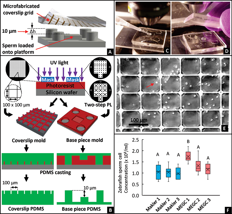
FIG. 1. Fabrication and use of a Microfabricated Enumeration Grid Chamber (MEGC) for concentration estimation of zebrafish sperm. (A) Overview of the grid-patterned coverslip with 100 × 100 µm squares placed on top of posts that create a 10-µm tall chamber to create a known volume (1 × 10⁻⁴ µl for 100 × 100 × 10 µm) for accurate estimation of sperm concentration. (B) The microfabrication process for the MEGC was photolithography and PDMS casting. The mold for the coverslip was prepared by exposing a spun negative photoresist masked by a 100 × 100 µm square grid pattern. The mold for the base piece was prepared by two-step photolithography, yielding a 10-µm height difference between the central platform and the adjacent pillars. For use of the MEGC, (C) zebrafish sperm samples were pipetted onto the central platform of the base piece, (D) followed by placing the coverslip on top. (E) To estimate sperm concentration, sperm samples were observed with a dark-field microscope (200-× magnification).